BESA Simulator Software


NEW NEW NEW
image-content-top_besa-simulator-overview

BESA Simulator

Create your own evoked and induced data

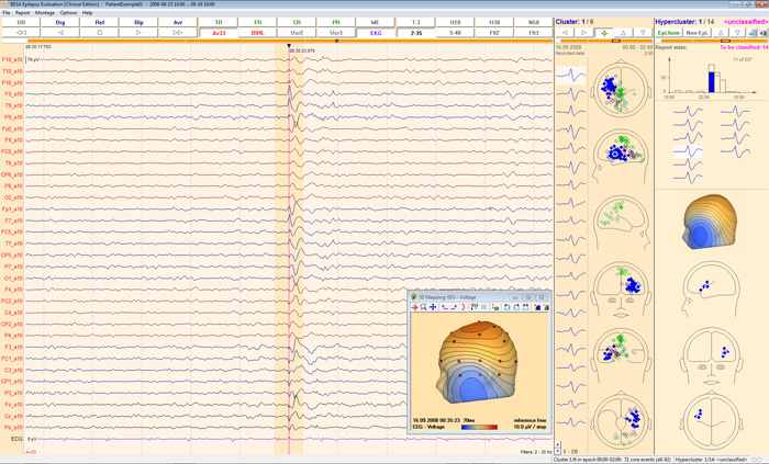
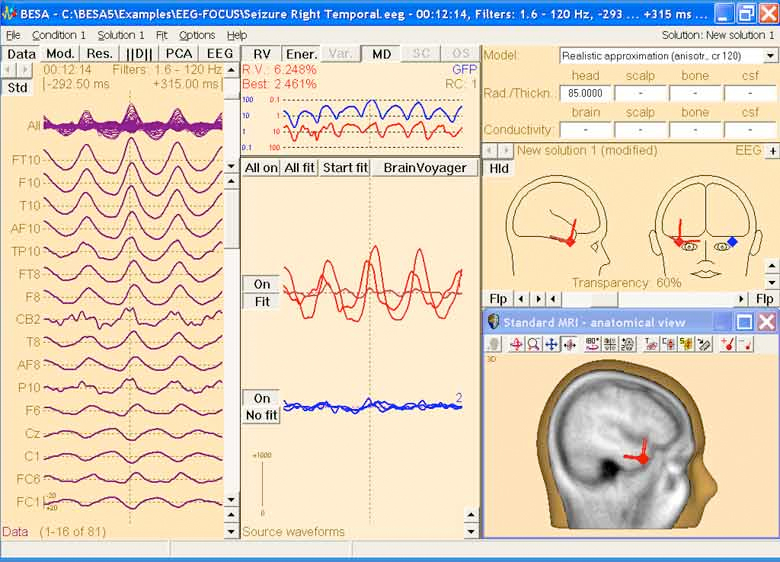
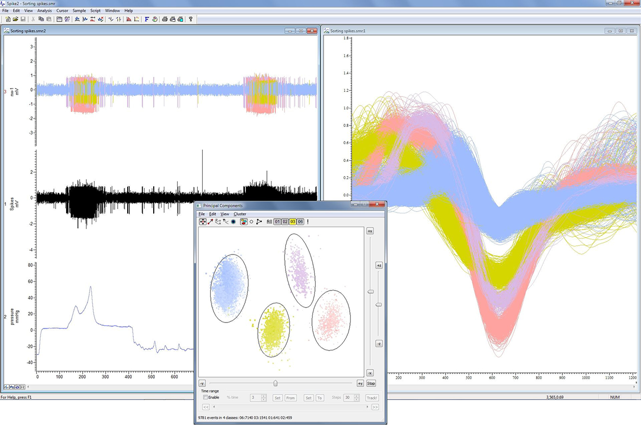
BESA Simulator is the perfect tool for simulating evoked and induced brain activity. This free software developed by Patrick Berg allows you to place your sources in the brain, assign them a time-course and save your data with an electrode layout of your choice. You can simulate average data or raw data choosing your own real dataset for overlaying the modeled activity. This allows simulating induced data, as well as evoked data. Use the simulated data to examine your source or coherence analysis techniques.

Do you manage to localize the simulated sources? Use BESA Simulator to create your own theories about brain networks underlying your data and see if they hold true in real data. BESA Simulator is an ideal instrument for learning and also teaching about induced and evoked brain activity!

With BESA Simulator you can:

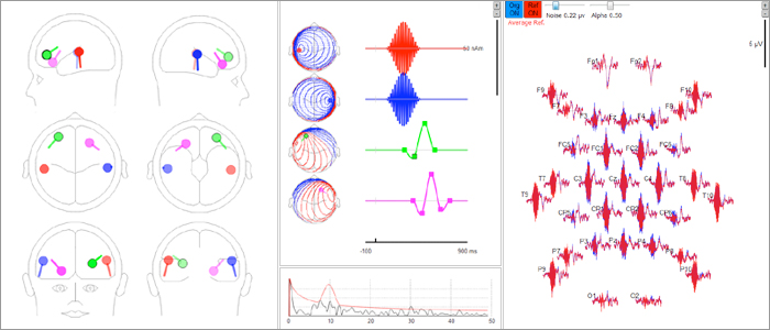
  • Simulate both EEG and MEG data
  • See maps resulting from a dipole anywhere within the head
  • Add any number of dipoles to a model
  • Generate independent waveforms for each dipole (source waveforms). You can also read in source waveforms from an ASCII file (BESA swf)
  • See the surface data resulting from the model as reference-free, average reference, Laplacian (CSD) reference, or using any electrode / sensor as reference
  • Place a cursor anywhere in the time interval and visualize the scalp activity at that time point
  • Specify the parameters of the spherical head model
  • Add coherent noise to the data defined in terms of rms level and level of alpha activity
  • Learn about filter effects by displaying the effects of applying various types of filters to your simulated data
  • Use any electrode or MEG sensor configuration. MEG configurations for 4-D, CTF, and Neuromag systems are supplied as examples.
  • Save and read the models you have generated
  • Save the generated data in BESA avr (ASCII) format
  • Simulated raw data can be saved in BESA Simple Binary (Generic) format.
  • Simulate activity using individual head models (FEM) generated by BESA MRI


Request a Quotation

BESA Epilepsy Software


image-content-top_besa-epilepsy-overview

BESA Epilepsy 1.0

CE certified software for fast review and clustering of epileptiform EEG activities

  • Run an automatic spike detection with 91% sensitivity in adults and children.
  • Spike detection takes at the most 3h on a 24h EEG.
  • Select a patient and the EEG review epoch instead of files.
  • Choose optimized montages and filters in EEG Review by 1-click.
  • Decide, if hyperclusters are epileptiform or not. Spend 5 minutes a day for each patient.
  • A final report is generated automatically. Add EEG images and comments manually by 1-click.
  • Manage the data of 12 different EEG data formats in one program.
  • Easy to install and quick integration into your system.
  • Learn the program very fast based on a tutorial including four example patients.
  • Available in Clinical Edition for countries accepting the CE certification for clinical use. Note that this edition is not FDA approved.
  • Available in Research Edition for research purposes only. This edition is also intended to be used in our new reliable pipeline to analyze interictal spikes and seizure onset in EEG and MEG.

You want to save time on long-term EEG evaluation?

BESA Epilepsy 1.0 provides automatic spike detection with 91% sensitivity in adults and children. The clustering and hyperclustering of the detected interictal activity is an efficient feature for the EEG evaluation. Spend 5 minutes a day to evaluate a 24h EEG. Print your final report or store it a PDF.

Start the spike detection directly after your recording starts and you can evaluate the EEG nearly online. For offline analysis BESA Epilepsy 1.0 requires approximately 3h to analyze a 24h EEG.

You want to evaluate EEGs recorded by different systems in one application?

BESA Epilepsy 1.0 is able to read 12 different EEG data formats. Manage your data in one common database. Select the patient and choose a review interval to start the EEG evaluation. Don’t think in files any longer.

  • BESA
  • Coherence/ITMed
  • Compumedics
  • EBNeuro
  • EDF+
  • EGI MFF
  • Grass-Telefactor (TwinRef)
  • Micromed
  • Nicolet Nervus
  • Nihon Kohden
  • Stellate Harmonie
  • XLTEK

    Spike detection

    Automated detection and fast clustering

    • Automated detection of epileptiform discharges, e.g. spikes, sharp waves, with 91% sensitivity in adults and children.
    • Automated detection using 29 regional brain sources. High sensitivity – also for spikes in fissures!
    • Detection takes at the most 3h on a 24h EEG.
    • Runs on routine EEG and LTM EEG.
    • Program execution recommended on each acquisition unit (Clinical Edition only) and/or server in “nearly-online” or offline mode.
    • Independent of acquisition software with additional review capability.
    • Easy, intuitive workflow to set up detection during EEG monitoring.

    image-content-top_besa-epilepsy-features-spike-detection

    Do you need to page through 4537 detections over a 24 h EEG?

    No. Since clustering and hyperclustering are done after spike detection.
    “Fast evaluation of interictal spikes in long-term EEG by hyper-clustering”


Request a Quotation

BESA Research Software

BESA Research 7.1

Choose the best signal processing for your EEG and MEG data

BESA is the most widely used software for source analysis and dipole localization in EEG and MEG research. BESA Research has been developed on the basis of 30 years experience in human brain research by the team around Michael Scherg, University of Heidelberg, and Patrick Berg, University of Konstanz.

BESA Research is a highly versatile and user-friendly Windows® program with optimized tools and scripts to preprocess raw or averaged data for source analysis and connectivity analysis. All important aspects of source analysis and source imaging are displayed in one window for immediate selection of a wide range of tools. The same holds true for the source coherence / time-frequency module, and other analysis windows. BESA Research provides fast and easy hypothesis testing, a variety of source analysis algorithms including cortical imaging and volume imaging methods, integration with MRI and fMRI, age-appropriate template head models (FEM) as well as the possibility to import individual head models (BEM and / or FEM) generated by BESA MRI.

Connectivity analysis, a unique feature for viewing brain activity

BESA Research transforms surface signals into brain activity using source montages derived from multiple source models or beamformer imaging. This allows displaying ongoing EEG/MEG, single epochs, and averages with much higher spatial resolution.


The BESA Connectivity solution and the Source Coherence Module provide an extremely fast and user-friendly implementation of time-frequency analysis based on wavelets and / or complex demodulation. Users can create event-related time-frequency displays of power, amplitude, or event-related (de)synchronization and several connectivity measures for the current montage using brain sources or surface channels. Induced and evoked activities can be separated. Source connectivity analysis reveals the functional connectivity between brain regions by reducing the volume conduction effects seen in surface coherence. Users can select a time-frequency window for (bilateral) beamformer and dynamic imaging of coherent sources (Gross et al., PNAS 2001).

More than just dipole source localization

BESA Research covers the whole range of signal processing and analysis from the acquired raw data to dynamic source images:

Data review and processing

Source montages and 3D whole-head mapping

ERP analysis and averaging

Source localization and source imaging

Individual MRI and fMRI integration with BESA MRI and BrainVoyager

Source coherence and time-frequency analysis


Request a Quotation

Cadwell Sierra Summit

The Sierra Summit is a powerful system for all of your neurodiagnostic monitoring

The Sierra Summit is the result of 40 years of research and development. Responding to customer feedback, new electrodiagnostic (EDX) technology, and broader learning, Cadwell delivers one of the most dynamic pieces of neurophysiology diagnostic equipment. In 2018, Cadwell became the first EMG company to offer fully integrated ultrasound. Today, Cadwell leads the charge in neurodiagnostic technology capabilities in EMG, nerve conduction, evoked potentials, ultrasound, and chemodenervation.

Customize the system you need with a variety of amplifiers, accessories, carts and test protocols

  • Cadwell’s most advanced amplifiers offer 1 to 12 channels of sophisticated filtering and noise reduction
  • Select from a variety of carts, computers and monitors, or provide your own
  • Protocol licensing allows you to select only the protocols that you need

The Sierra software offers features developed to improve accuracy and reduce testing time. The smart and flexible user interface can fit your workflow and expand your capabilities.

NCS – Sierra Summit allows you to perform an entire EMG or NCS from the palm of your hand with the new and improved StimTroller Plus™

Control intensity, pulse width, distance, nerve selection, site selection, trace  selection, and more with the StimTroller Plus. Adjust the angle and distance between probes or remove them to attach  electrodes, needles or other stimulators. Engineered with reinforced housing and stainless steel mechanics, the StimTroller Plus will help you deliver efficient EDX testing experiences.

Intuitive and Sophisticated Software, rich with features designed to improve accuracy and reduce testing time.

EMG – Maximize clinical information reducing testing time

Increase confidence in your diagnosis with a seamless transition to live quantitative EMG.

Ultrasound

Sierra Summit™ with ultrasound is a complete integrated electrodiagnostic and imaging solution. Two ultrasound options with a variety of transducers are available to fit your needs and your budget.

EP – Evoked Potentials

Sierra Summits is a smart, flexible and efficient EP system that offers cutting edge features.

Multi-Modality: Complete neurodiagnostic solutions

EEG, EMG, NCS, EP and Ultrasound. Cadwell EDX solutions are engineered to work on a single cart or as separate systems connected with CadLink® data management.

Reporting

The Sierra Summit™ is designed with many timesaving and intuitive reporting features: Quick report, tab data summary view, auto findings composer, custom reports, anatomy view and EMR integration and data management.

Learn more:

Sierra Summit Advancing Clinical Neurophysiology

Sierra Summit EMG-NCS Brochure

Sierra Summit Evoked Potentials Flyer

Sierra Summit Family Brochure

Sierra Summit Ultrasound Flyer

Symbiotic Devices Carts Selection Catalogue

The Case for Neuromuscular Ultrasound – Whitepaper


Request a Quotation

NeuroConn DC-STIMULATOR MC

Programmable multi channel stimulator for neuroscientific research

The DC-STIMULATOR MC is a stimulator for use in scientific research. It provides a stimulation with weak direct or alternating current (transcranial electrical stimulation – tES) within non-invasive Neurophysiology. The electrical charge and current density applied are far below the threshold for releasing a stimulus. Depending on the duration, the used current, the current density and the frequency the stimulation either activates or inhibits cortical activity. It has therefore a modular effect on existing neuronal elements.

Its main focus of application is in clinical research into multi-channel, controlled, supervised and simultaneous tDCS / tACS / tRNS stimulation of patients and patient groups.

The DC-STIMULATOR MC provides up to 16 electrically independent, freely programmable, micro-processor-controlled constant current sources using independent channels. It meets the highest safety standards thanks to (hardware- and software-based) multistage monitoring of the current path. By continuously monitoring electrode impedance it can accurately maintain the user-defined current amplitude while also detecting if there is insufficient contact with the skin, which will then automatically terminate stimulation to ensure subject safety.

The DC-STIMULATOR MC can also be used during functional magnetic resonance imaging (fMRI) and, in addition, can be combined with the NEURO PRAX TMS/tES allowing full-band DC-EEG to be recorded during multi-channel tES.

Learn more:

DC-Stimulator MC Brochure


Request a Quotation

Signal software

Signal Software V7

Signal is a sweep based data acquisition and analysis package. Its uses range from a simple storage oscilloscope to complex applications requiring stimulus generation, data capture, control of external equipment and custom analysis. This flexibility makes it ideal for a wide range of applications including transient capture, patch and voltage clamp, LTP studies, evoked response and TMS.

  • Signal is simple to set up for data capture and analysis
  • Signal has the sampling and analysis features most researchers need, in a user-friendly environment. A built-in script language automates repetitive tasks and provides additional tools for custom analyses and applications.
  • Signal includes functions for specific application areas including dynamic clamp, whole cell and patch clamp electrophysiology and evoked response including control of magnetic and other stimulus devices.
  • Signal imports data recorded by many other systems, you can take advantage of this extremely versatile system to analyse existing data.
  • Optional windows can show dynamic information in text and images at large scale, to be visible across the lab.

The powerful data capture and time-saving analysis functions of Signal together with one of the CED 1401 family of laboratory interfaces make an extremely flexible and cost effective addition to any laboratory.

Learn more:

CED Signal Software Brochure

CED Signal Software website


Request a Quotation

Spike 2 Software

Spike 2 Software, Version 10

Spike2 is a multi-channel continuous data acquisition and analysis package. Uses range from a simple chart recorder to complex applications requiring stimulus generation, data capture, scrolling or triggered displays, control of external equipment and custom analysis. This flexibility makes it suitable for a wide range of applications for specific analysis tasks.

  • Spike2 is ideal for applications in many fields including electrophysiology, neurophysiology, sports science, pharmacology and many more.
  • Spike2 provides power and flexibility for demanding applications, such as extracellular multi-electrode recording and complex stimulus timing.
  • Spike2 features a built-in script language to customise the program and provide control from simple automation to addition of complex analyses.
  • Spike2 imports data recorded by many other systems, you can take advantage of this extremely versatile software to analyse existing data.
  • Spike2 can export data to spreadsheet, text, binary and MATLAB® files.
  • Spike2 is continually updated; the latest version adds new features for free throughout the life cycle of the version.

These powerful data capture and analysis functions, together with one of the CED1401 family of laboratory interfaces, make Spike2 an extremely flexible, cost-effective and time-saving addition to any laboratory.

Learn more:

Spike2 Software Brochure

Spike2 Software supplier website


Request a Quotation

CED Whisker Stimulator 3901

Controls an array of piezo-electric stimulator elements

The CED 3901 whisker stimulator is a controller for an array of piezo-electric stimulator elements, offering precisely controlled temporal patterns, with repeatable stimulation. Each 3901 drives 9 stimulator elements and up to 7 can be linked, to control up to 63 elements.

It is mains-powered and is controlled by 8 bits of the digital output of a CED 1401, or by USB. Each of the stimulator elements can be connected to one of four sources; either of the two external waveform inputs, an individual programmable pseudo-random noise generator, or mute.

Software control is provided as a demonstration Spike2 script.

  • Precisely control temporal patterns with repeatable stimulation
  • Horizontal, vertical and diagonal control
  • 9 to 63 output channels ±45v @ 10mA
  • Drive grids of 3×3 up to 7×9
  • Filtered outputs 2nd order low pass Butterworth (1kHz)
  • Output muting
  • Independent pseudo random noise generators
  • Adjustable noise levels
  • Use with Micro or Power intelligent interface or driven from other DAQ system
  • Software controlled – USB or 8-bit digital communication
  • Size – Micro1401 format: 366 x 48 x 217mm (14.4 x 1.75 x 8.5 ins) (W x H x D)

Find out more

Request a Quotation

CED 4001-16 Mains Pulser

The CED 4001-16 Mains Pulser generates mains-frequency-locked pulses that can be used to reduce the magnitude of mains pickup (‘hum’) in recorded signals. The output is on a BNC connector, double-isolated from mains and consists of a train of 150 microsecond positive-going 5V TTL pulses, suitable for direct connection to a 1401 Event input.

The Mains Pulser will run off any mains supply between 100 -240 V AC, 50 – 60 Hz. The case is earthed to mains earth. Current consumption is less than 100 mA.

Box size: 104mm x 126mm x 53mm (W x D x H).

Find out more


Request a Quotation

CED 4003 Potentiometer

The CED 4003 is a simple linear 10k potentiometer, intended to provide manually adjustable signal division. It is cased and has BNC leads that allow input connection from a voltage source such as a 1401 DAC waveform output, with an output connection suitable for an ADC waveform input.

The potentiometer output is controlled by a slider, with markings and writeable labels.

Case size: 166mm x 70mm x 28mm, lead length 2 metres.

The input voltage should not exceed 30V.

Found out more


Request a Quotation

CED 3505 Programmable Attenuator

The CED 3505 and 3505HP are single-channel programmable attenuators offering main attenuations from 0dB to over 100dB in 2.5, 3 or 5dB steps.

Control of the attenuation and other programmable features of the 3505 is via an internal USB-serial port. It may be controlled by scripts in Spike2 or Signal. Fast attenuation changes may also be implemented via a parallel port designed to interface with a CED1401 digital output. In this way, the sequencer facility in the CED Spike2 software package can control the attenuation for real-time synchronisation under experimental conditions.

The standard 3505 has an additional output connector producing an “attenuation changed” pulse. The jack socket carries the output from an internal audiometric headphone amplifier, designed to drive standard audiometric headphones to sound pressure levels of around 140dB. The attenuator output signal can be routed to one or both headphones, and there is an independent calibration attenuator for each headphone channel.

3505 programmable-attenuator front

3505 programmable-attenuator rear


Box size: 240mm x 240mm x 46mm (W x D x H), external mains-driven power supply.

Find out more

Owners Handbook


Request a Quotation

CED 2502SA

Skin Conductance Unit

The 2502SA stand-alone skin conductance unit is designed to take direct measurements of the conductivity between two skin electrodes by maintaining a 500mv DC potential across them, and then measuring the current needed to maintain this potential. This current value is then represented and output as a scaled voltage, suitable for connection to a CED 1401 laboratory interface.

The 2502SA uses the constant-voltage principle and has manually switched conductance ranges of 10, 25 and 100 micromhos. The sockets accept 1.5 or 2 mm plugs.

The CED 2502SA is offered as research equipment and is not sold as a medical device within the meaning of the European Medical Device Directive.

2502SA CED Skin Conductance Unit

Find out more


Request a Quotation

CED Power1401-3A

High-performance data acquisition interface

The Power1401-3A is our latest high-performance data acquisition interface. It uses advanced processor technology to give you all the power and flexibility you need in a state-of-the-art laboratory interface.

Fast data acquisition and analysis

The Power1401-3A records waveform data, digital (event) and marker information, and can generate waveform and digital outputs simultaneously for real-time, multi-tasking experiment control. The Power1401 has a 1 GHz Marvell processor (essentially an ARM V5TE core) and up to 2GB of on-board memory, to facilitate high speed data capture, independent waveform sample rates and complex on-line analysis, freeing valuable time for the host computer to perform other tasks, such as data manipulation and further analysis.

Find out more


Request a Quotation

AlgoMed

Pressure Algometer

The AlgoMed is the first software-based computerized Pressure Algometer to offer real-time visual & auditory feedback to control & monitor applied pressure rates to measure pressure pain threshold.
Algometers are designed to quantify and document levels of tenderness via pressure pain threshold measurement and pain sensitivity via pressure pain tolerance measurement. Pressure algometry is a reliable measure of pain in muscles, fascia, joints, tendons, ligaments and the periosteum.
The challenge in performing pressure algometry tests is to apply continuously ascending pressure at a constant rate on the patient’s selected body site without having the possibility to monitor it in real time.

Features:

  • Real-time visual & auditory feedback
  • Comprehensive test statistics
  • Body site selection
  • Customizable outcome-based color graphic reports
  • Adjustable protocols, including thresholds and Ramp & hold

AlgoMed_Medoc


Find out more Product Brochure

Medoc Quantitative Sensory Testing –  Solutions for Neurophysiology


Request a Quotation

Q- Sense

Simple, validated, thermal testing for small peripheral nerve fibers

Q-Sense is a portable, easy-to-use and affordable quantitative sensory testing (QST) device for clinical, research and pharmacologic trial use. Q-Sense offers scientifically validated measures of warm, cool and heat-pain thermal sensory thresholds. All of these can be useful determinants in the evaluation of peripheral sensory nerve function, neuropathic pain, diabetic, chemotherapeutic, and other small-fiber neuropathies.

Key features:

  • Sensitive and reproducible
  • Pre-programmed test algorithms
  • Versatile patient database
  • Normative reference data
  • Easy-to-interpret clinical test report
  • Easy data export to excel

Q-Sense Medoc

Q-Sense-thermode

Q-sense Symbiotic Devices

Learn more:

Medoc Q-Sense Product Brochure

Medoc Quantitative Sensory Testing –  Solutions for Neurophysiology

Medoc TSA2, Q-Sense and TSA-Air2 Comparison flyer


Request a Quotation

NeuroConn DC-STIMULATOR MR

Stimulator for Neuroscientific Research

The DC-STIMULATOR MR is an fMRI-compatible stimulator for use in scientific research. It is an extension of our single-channel DC-STIMULATOR PLUS, so it benefits from multi-stage monitoring and complete range of tES modes of operation: transcranial direct current simulation (tDCS), transcranial alternating current stimulation (tACS) and transcranial random noise stimulation (tRNS).

The DC-STIMULATOR MR allows to apply transcranial electrical stimulation (tES) during functional magnetic resonance imaging (fMRI) to localize the exact position of cortical activation.

Features:

  • 1 channel, unipolar (DC) and bipolar (AC) stimulation
  • Adjustable range of current ±4,500 µA, maximum adjustable current strength ±1,500 µA (peak-to-peak)
  • Adjustable application time up to 30 minutes
  • Frequencies adjustable up to 250 Hz, phase freely adjustable
  • 4 standard modes – single (continuous stimulation) – pulse (cyclical stimulation activation/deactivation) – sinus (sinus wave) – noise (normally distributed)
  • Use during fMRI, no interference of the MRI images during EPI sequence
  • RF filter module with MRI-compatible cables and electrodes
  • Customer-specific programs possible (optional)
  • “Study mode” for blind processing of genuine and ‘pseudo’ stimulation (optional)
  • External trigger input and output (optional)

Learn more

NeuroConn DC-STIMULATOR MR Brochure

DC-STIMULATOR MR AppNote Single Channel fMRI-tES

DC-STIMULATOR MR AppNote Dual Channel fMRI-tES


Request a Quotation

NeuroConn DC-STIMULATOR

DC Stimulator for cranial electrotherapy and neuroscientific research

The DC-STIMULATOR is a stimulator for cranial electrotherapy that provides a stimulation using weak direct current (transcranial Direct Current Stimulation tDCS) within non-invasive interventional neurophysiology. The electrical charge and current density applied are far below the threshold for releasing a stimulus and have a modular effect on existing neuronal elements.

Depending on the duration, used current, current density and frequency, the stimulation is effective on inhibiting or activating cortical activity.

The DC-STIMULATOR is used for transcranial Direct Current Stimulation for symptom relief in depression.

NeuroConn stimulators offer CE-certification and TGA certification, multi-stage monitoring and complete range of tES modes of operation:
tDCS – trans-cranial direct current stimulation
tACS – trans-cranial alternating current stimulation
tRNS – trans-cranial random noise stimulation

These currents influence neurotransmitter channels by changing the electrical charge in nerve cell membranes. In turn this changes the excitability of brain cells.

Studies have shown that tDCS can be used as a therapy method for various brain disorders that would otherwise be treated with therapeutic drugs, often associated with harmful side effects.

The NeuroConn DC stimulator and NeuroConn DC Plus offer 1 channel (anodal and cathodal) trans-cranial stimulation, adjustable application time of up to 30min with adjustable current up to 4.5mA is also included. The neuroConn trans-Direct Current Stimulator (tDCS) is a microprocessor-controlled constant current stimulator. It offers several safe and reliable stimulation modes that can be programmed.

In addition the neuroConn system offers extra software options including an advanced sham feature allowing double blind trials and a SIMPLE mode that restricts the use of the stimulator allowing the user to only choose from 4 predefined settings.

Learn more:

neuroCare DC-Stimulator Brochure

neuroCare tDCS application

neuroConn tDCS Effectiveness Studies


Request a Quotation

TriggerBox / TriggerBox Extension

The TriggerBox helps to handle and merge triggers arriving from different sources, which is quite often a requirement in complex laboratory setups.

TriggerBox

  • TriggerBox has connectors for parallel/LPT as well as BNC inputs; the latter are all isolated from each other as well as from the other input connectors.
    It is equipped with 8 toggle switches to regulate bit-wise which input should determine the value of the output parallel port, where the EEG amplifier is connected.
  • It can provide connection to the 16th bit of the BrainAmp trigger input, via an optical fibre cable or BNC connector.
  • It offers a stretching function for the 8th and 16th bits which is useful if the incoming TTL pulses are too short for the amplifier and would otherwise go undetected.
  • It is powered by USB.
  • New as of revision 2 – TriggerBox is now even more flexible and provides a virtual serial port via USB that can be directly addressed from your stimulus presentation software.

TriggerBox Extension

  • TriggerBox Extension offers 8 additional BNC connectors which can be linked to the 8 upper bits (from lines 8 and 15) of the BrainAmp or used as a standalone solution for amplifiers such as actiCHamp or V-Amp in situations where stretching, switching and isolation functions are not required.
  • It can be connected to the output trigger port of the actiCHamp in order to lead the outgoing signal to the BNC ports.

Learn more:

SyncBox Brain Products Website

Press Release: TriggerBox 2 – millisecond precise trigger without a parallel port


Request a Quotation