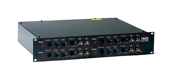
The TriggerBox helps to handle and merge triggers arriving from different sources, which is quite often a requirement in complex laboratory setups.
TriggerBox
- TriggerBox has connectors for parallel/LPT as well as BNC inputs; the latter are all isolated from each other as well as from the other input connectors.
It is equipped with 8 toggle switches to regulate bit-wise which input should determine the value of the output parallel port, where the EEG amplifier is connected. - It can provide connection to the 16th bit of the BrainAmp trigger input, via an optical fibre cable or BNC connector.
- It offers a stretching function for the 8th and 16th bits which is useful if the incoming TTL pulses are too short for the amplifier and would otherwise go undetected.
- It is powered by USB.
- New as of revision 2 – TriggerBox is now even more flexible and provides a virtual serial port via USB that can be directly addressed from your stimulus presentation software.
TriggerBox Extension
- TriggerBox Extension offers 8 additional BNC connectors which can be linked to the 8 upper bits (from lines 8 and 15) of the BrainAmp or used as a standalone solution for amplifiers such as actiCHamp or V-Amp in situations where stretching, switching and isolation functions are not required.
- It can be connected to the output trigger port of the actiCHamp in order to lead the outgoing signal to the BNC ports.

Learn more:
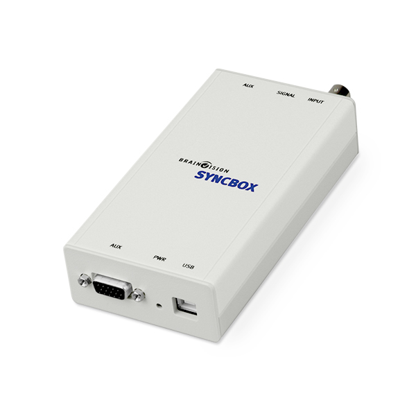
SyncBox Brain Products Website
Press Release: TriggerBox 2 – millisecond precise trigger without a parallel port
Request a Quotation |


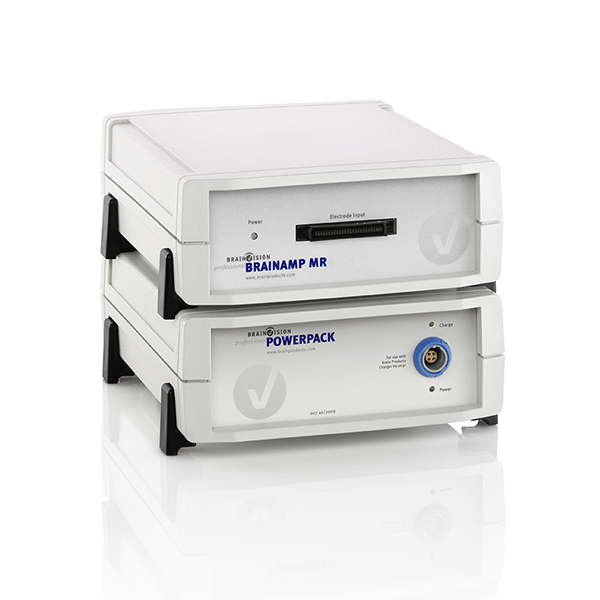
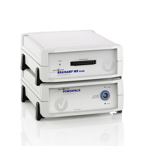 The BrainAmp MR plus enhances the already outstanding features of the