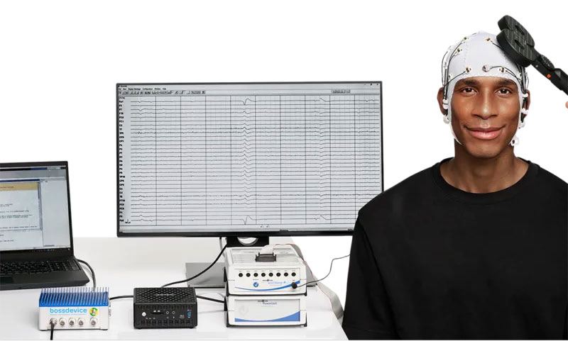
STE Compact for LiveAmp:

-
Enhances mobile configurations by incorporating physiological data and trigger choices
-
Integrates 6 auxiliary sensor ports along with a dedicated connection for pulse oximetry
-
Permits both wireless and 8-bit wired trigger setups
-
Offers versatile power configurations suited for lightweight arrangements or extended recording sessions
Overview
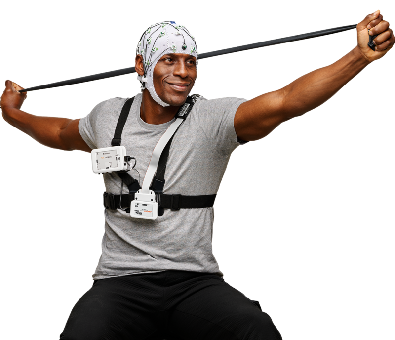
The STE compact (Sensor and Trigger Extension) is a lightweight extension module for the LiveAmp amplifier. It allows researchers to add physiological sensors and trigger capabilities to mobile EEG setups. Utilizing the LiveAmp’s internal memory, data recording is entirely independent of a computer.

Key Features
Sensors and Inputs
-
Auxiliary (AUX) Inputs: Features 6 AUX ports compatible with all Brain Products sensors (such as Photo Sensors for display timing, Force Sensors for heel strikes, Respiration Belts, or GSR Sensors).
-
Dedicated Digital Input: Designed for a Pulse Oximetry Sensor to record blood oxygen saturation (SpO2), heart rate (HR), and photoplethysmogram (PPG).
Triggering Capabilities
-
Wireless Triggering: Includes a built-in Wireless Trigger Receiver that pairs with a transmitter. It delivers low-latency (~2.5 ms) and reliable (+/- 1 sample) transmissions up to a 10-meter distance.
-
Hyperscanning Support: A single Trigger Transmitter can connect to up to 5 STE compact receivers simultaneously.
-
Wired Triggering: Supports an 8-bit wired trigger connection via a 9-pin D-Sub connector.
Power Options and Portability
-
Attachment: Features a built-in belt clip for easy mounting to the subject or the LiveAmp Mobility Set.
-
Flexible Power Supply: Powered by internal, exchangeable AA batteries (up to 10 hours of runtime). It supports “hot-swapping” to replace batteries during recording without interruption. It can also run directly off the LiveAmp’s power to minimize weight, or use an external power bank to extend runtime.

Getting started
STE compact for LiveAmp
How to set up STE compact & LiveAmp step by step


 Know the specs:
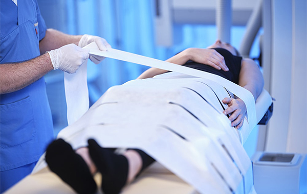
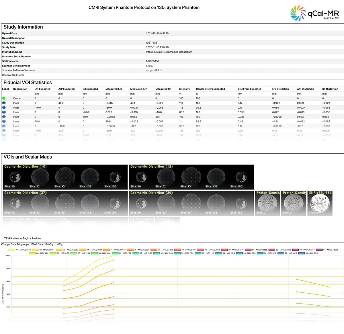
 Evaluated MRI Characteristics
 LabMaestro Simulator Software Brochure
 Learn more: