
EEGFocus combines digital EEG review with advanced analysis features such as 3D whole head mapping, brain source montages and images, spike pattern search and averaging, spectral analysis, DSA trend analysis, on-line correction of eye and EKG artifacts etc.
Digital EEG Review
EEGFocus provides a user-friendly interface for immediate analysis of abnormal patterns during review, e.g. spikes or rhythmic EEG activities. Similar patterns can be searched for in the whole EEG and averaged, for example, to analyze a spike or seizure onset.
- Ergonomic buttons and key functions for fast manual or automatic paging. Overlapped paging to detect EEG abnormalities at page boundaries. Easy tool to measure peaks and frequencies
- Video synchronized paging, optionally with whole head mapping
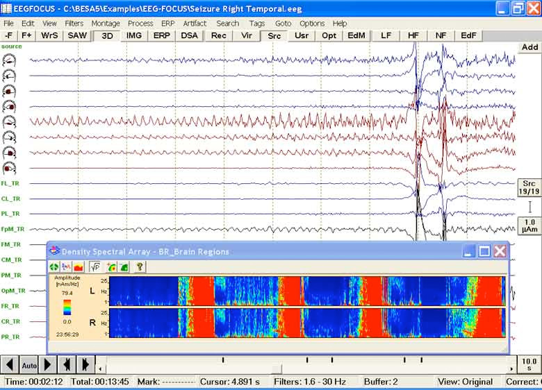
- Fast navigation using the event bar or the DSA trend display with separation of the hemispheres (DSA = density spectral arrays)
- Advanced digital filters including zero-phase shift, band and notch filters
All-in-one software for fast review and advanced EEG analysis
3D whole head mapping
Whole head maps provide a complete overview and a better interpretation of the topography of EEG abnormalities. Just click on a detected pattern to obtain 3D whole head maps. Click on a particular view to obtain a series of maps showing the evolution over time.
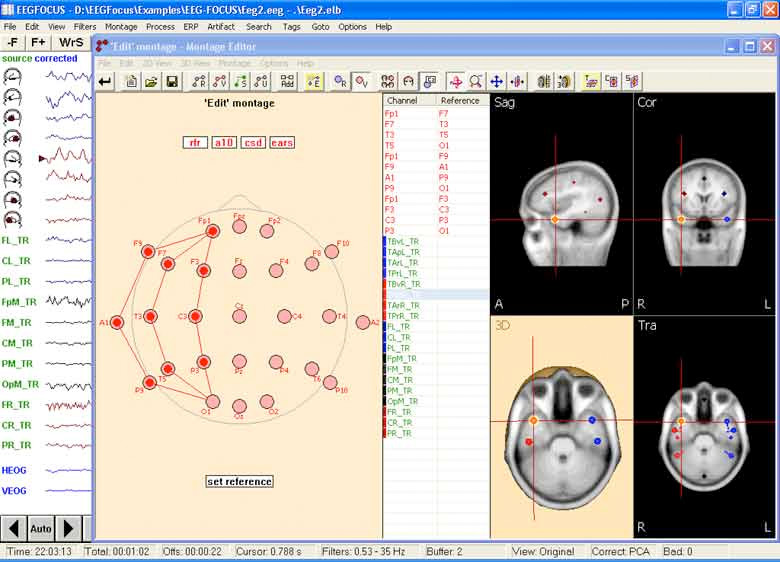
Source montages
Conventional montages, e.g. longitudinal bipolar, measure potential differences at the scalp. EEGFocus uses all electrodes and multiple sources to transform the scalp EEG into the brain. Thus, activities generated in the right and left hemisphere, or in different brain regions, can be separated to a large degree. This leads to a better visibility of focal abnormalities in the source waveforms and separation of the on-going EEG rhythms.
Spectral analysis
Spectral analysis can be done with high frequency resolution over a marked epoch, e.g. at seizure onset, or over the whole EEG. DSA trend analysis provides a fast overview of the spectral power of the whole EEG. By using brain source montages for DSA, hemispheric or regional differences can be enhanced, e.g. to reveal focal seizure activity.

Pattern search
After identifying a pattern of interest, e.g. a spike, the marked signal can be used as a template to search the whole EEG for repeated occurrences of this pattern. The search can be based on a single channel or on the spatio-temporal pattern across the current montage. In combination with brain source montages recurrent focal activities can be detected reliably.

Artifact correction and ERP
EEGFocus uses all EEG and EKG channels in a unique automated tool to detect and correct eye and EKG artifacts.

Request a Quotation |
